Medical Image Analysis
Home
About Us
People
Teaching
Research
Publications
Awards
Links
Contact
Internal
Medical image analysis is one of the main application fields for
novel digital image processing methods. We have explored a number
of applications that serve as demonstrators for our methods for
image denoising, structure enhancement, segmentation and classification.
Often this is done in close collaboration with medical experts.
-
Segmentation of Magnetic Resonance Images
Nonlinear diffusion scale-spaces have been introduced into the so-called hyperstack, a segmentation method that exploits the deep structure in scale-spaces. Evaluation on clinical imagery demonstrates the advantages of this approach. Different two- and three-dimensional medical applications are studied in [1], [2], [3], [4]. -
Multiscale Representation of Trabecular Bone Structures
The trabelucar structures of bones are responsible for the stability of the bones with respect to external forces. Their density and orientation at multiple scales gives important diagnostic information for patients suffering from osteoporosis. In order to obtain such a mutiscale representation, three-dimensional variants of coherence-enhancing anisotropic diffusion filters have been developed [5], [6]. -
Visualisation and Evaluation of Vessel Structures in
3-D Rotational Angiography
Three-dimensional rotational angiography is a powerful imaging techniques for vascular structures, but suffers from noise. We have evaluated a number of linear and nonlinear diffusion techniques for denoising these data sets by means of in vitro experiments [7], [8]. -
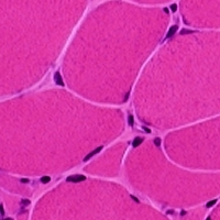
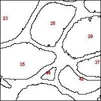
Analysis of Muscle Fibres
In order to assess neuromuscular diseases, it is useful to have precise meaurements of the muscle fibre size and its distribution. We have proposed a fully automated method that segments muscle fibres using edge- and region-based active contours. Multiple morphometric parameters are extracted. Comparisons with manual measurements by experts demonstrate a high rate (98 percent) of correct classifications [9], [10]. The images below depict a muscle fibre image and a corresponding segmentation.
-
Fast Kinematic MR Imaging of the Eye and Orbit
For assessing patients with restricted eye movement, fast kinematic magnetic resonance (MR) imaging can be used. Since the data are degraded by noise, preprocessing using anisotropic diffusion filtering has been applied. The results demonstrate that an almost fluent visualisation of the eye and orbital dynamics can be obtained [11]. -
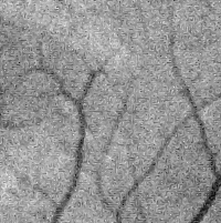
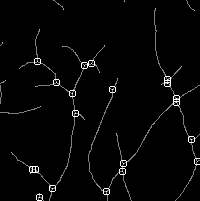
Retinal Vessel Detection Using the Local Radon Transform
The analysis of retinal blood vessels provides important diagnostic information. For the automatic detection of retinal blood vessels, we have developed a system that is based on local Radon kernels and offers real-time capabilities. Comparisons with other methods on standard databases demonstrate the good performance of our approach [12]. Below is a noisy original image and the extracted vessel skeleton and its branching points.
-
Diffusion Tensor MR Imaging
Diffusion Tensor MR Imaging (DT-MRI) measures the diffusive properties of water molecules in the brain (and other tissues). Such information can be useful for connectivity analysis and assessment of stroke, schizophrenia and other diseases. DT-MRI creates matrix-valued data sets, so-called tensor fields. Our group has developed many methods for denoising, enhancement, segmentation, interpolatation and registration of tensor fields. These results are described in more detail on our tensor web page.
-
W. J. Niessen, K. L. Vincken, J. Weickert, M. A. Viergever,
Nonlinear multiscale representations for image segmentation,
Computer Vision and Image Understanding, Vol. 66, 233-245, 1997.
[Abstract] -
W. J. Niessen, K. L. Vincken, J. Weickert, M. A. Viergever:
Three-dimensional MR brain segmentation.
Proc. Sixth Int. Conf. on Computer Vision (ICCV '98, Bombay, Jan. 4-7, 1998), 53-58, 1998.- W. J. Niessen, K. L. Vincken, J. Weickert, B. M. ter Haar Romeny, M. A. Viergever:
Multiscale segmentation of three-dimensional MR brain images,
International Journal of Computer Vision, Vol. 31, 185-202, 1999.
- W. J. Niessen, K. L. Vincken, J. Weickert, M. A. Viergever:
Multiscale segmentation of volumetric MR brain images.
In H. Yan (Ed.): Signal Processing for Magnetic Resonance Imaging and Spectroscopy, Marcel Dekker, New York, 209-238, 2002.- J. Weickert, B. M. ter Haar Romeny, A. Lopez, W. J. van Enk:
Orientation analysis by coherence-enhancing diffusion.
Proc. Symposium on Real World Computing (RWC '97, Tokyo, Jan. 29-31, 1997), 96-103, 1997.
- J. Weickert:
Coherence-enhancing diffusion filtering,
International Journal of Computer Vision, Vol. 31, 111-127, 1999.
- E. Meijering, W. Niessen, J. Weickert, M. Viergever:
Evaluation of diffusion techniques for improved vessel visualization and quantification in three-dimensional rotational angiography.
W. J. Niessen and M. A. Viergever (Eds.): Medical Image Computing and Computer-Assisted Intervention - MICCAI 2001. Lecture Notes in Computer Science, Vol. 2208, Springer, Berlin, 177-185, 2001.- E. Meijering, W. Niessen, J. Weickert, M. Viergever,
Diffusion-enhanced visualization and quantification of vascular anomalies in three-dimensional rotational angiography: results of an in-vitro evaluation,
Medical Image Analysis, Vol. 6, No. 3, 217-235, September 2002.
Preprint.- T. Brox, Y.-J. Kim, J. Weickert, W. Feiden:
Fully-automated analysis of muscle fiber images with combined region and edge based active contours.
In H. Handels, J. Ehrhardt, A. Horsch, H. P. Meinzer, T. Tolxdorff (Eds.): Bildverarbeitung in der Medizin. Springer, Berlin, 86-90, 2006.- Y.-J. Kim, T. Brox, W. Feiden, J. Weickert:
Fully automated segmentation and morphometrical analysis of muscle fibre images.
Cytometry: Part A, Vol. 71A, No. 1, 8-15, 2007.
Revised version of Technical Report No. 177, Department of Mathematics, Saarland University, Saarbrücken, Germany, July 2006.- E. P. Stuijfzand, M. D. Abrŕmoff, K. J. Zuiderveld, L. M. P. Ramos, J. Weickert, M. P. Mourits, F. W. Zonneveld, W. P. T. H. Mali,
Fast kinematic MR imaging of the eye and orbit,
RSNA Electronic Journal, Vol. 1, 1997.- M. Krause, R. M. Alles, B. Burgeth, J. Weickert:
Fast retinal vessel analysis.
Journal of Real Time Image Processing, in press.
Revised version of Technical Report No. 320, Department of Mathematics, Saarland University, Saarbrücken, Germany, Dec. 2012. - W. J. Niessen, K. L. Vincken, J. Weickert, B. M. ter Haar Romeny, M. A. Viergever:
MIA Group
©2001-2023
The author is not
responsible for
the content of
external pages.
Imprint -
Data protection